The 4 Types of Neurons Running Your Entire Nervous System
By Tristan Siokos · Founder, Recalibrate · June 4, 2026
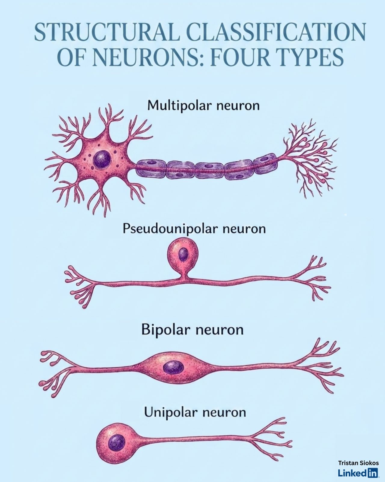
Most people think of a neuron as one shape. There are actually four. Each one is built differently because each one does a different job, and the architecture of each cell tells you exactly what kind of work it was made for. Here is what they are and what they actually do for you.
Architecture First: Why Shape Tells You Everything
Your nervous system contains roughly 86 billion neurons. They are not all the same shape. And the shape is not arbitrary.
Each neuron type is built the way it is because of the job it has to do. The number and arrangement of its processes, the length of its axon, the density of its dendritic tree, the location of its cell body relative to its inputs and outputs, all of these are structural solutions to specific functional problems.
When you understand the four structural types, you understand why your nervous system is built the way it is. Not as a list of facts to memorise, but as engineering you can read.
The four structural categories are: multipolar, pseudounipolar, bipolar, and unipolar. Each one appears in a specific location in the nervous system, does a specific category of work, and has a distinct evolutionary and developmental story.
Shape is the first lens. Function, neurotransmitter identity, and molecular signature add the rest. But start with shape. It tells you more than it appears to.
Multipolar Neurons: The Architecture of Cognition and Action
The multipolar neuron is the most abundant neuron type in the human nervous system. One long axon for output. Multiple dendrites extending from the cell body for input. The cell body sits in the middle, integrating everything it receives and deciding what to send.
The variety within the multipolar category is enormous.
Pyramidal cells are multipolar neurons in the cerebral cortex. Their apical dendrites reach upward toward the cortical surface, gathering input from across cortical layers. Their axons project long distances, forming the corticospinal tract that connects your motor cortex to your spinal cord. When you decide to move your hand, pyramidal cell axons are carrying that signal. When you think a complex thought, pyramidal cells in your prefrontal cortex are integrating thousands of inputs to compute the output. The cognitive architecture of your brain is built from pyramidal cells.
Purkinje cells are multipolar neurons in the cerebellum, among the most structurally complex cells in the entire nervous system. A single Purkinje cell may receive direct input from up to 200,000 other neurons via its elaborate dendritic arbour. They are the primary output neurons of the cerebellar cortex, critical for motor learning, timing, coordination, and balance. Every smooth, learned movement you make, from playing an instrument to walking without thinking, depends on Purkinje cell computation.
Motor neurons (lower motor neurons) are multipolar neurons in the spinal cord's anterior horn. Their axons exit the spinal cord and travel to muscles. Every voluntary contraction of every muscle in your body begins with a motor neuron firing. These cells are the final common pathway between your nervous system and your physical action in the world. They are lost in ALS and their loss explains that condition's progression.
The multipolar architecture, one output channel and many input channels, is the architecture of integration. These neurons are built to receive, weigh, and respond.
Pseudounipolar Neurons: The Sentinels of Your Body's Periphery
The pseudounipolar neuron has one of the most elegant developmental stories in neuroscience.
It starts life as a bipolar neuron: one process toward the periphery, one toward the central nervous system. During development, the two processes fuse near the cell body into a single trunk. That trunk then divides into two branches heading in opposite directions. The cell body sits to the side, off the main line of signal transmission. Structurally, it looks like one process with a cell body bubbling off it. Functionally, it is a through-line from your skin to your spinal cord.
Where they live: The dorsal root ganglia (DRG) clusters of cell bodies just outside the spinal cord, and the trigeminal ganglion for the face and head.
What they do: One branch extends into your peripheral tissues: skin, joints, muscles, and viscera. The other branch enters the spinal cord's dorsal horn. When that peripheral branch detects a stimulus, the signal travels straight through the cell body to the central branch and into the spinal cord. No synapse at the cell body. The cell body is a metabolic support station, not a processing node.
These neurons carry touch, temperature, proprioception (joint position and movement), and pain. Every time you feel a surface with your fingertip, know where your arm is without looking, or feel pain from a stubbed toe, pseudounipolar neurons are the first link in the chain from body to brain.
In the context of chronic pain: pseudounipolar neurons in the DRG are a primary target for peripheral sensitisation. When peripheral nociceptors lower their firing thresholds, it is often because ion channels and receptors on pseudounipolar axons have been altered by local inflammation, nerve growth factors, or ongoing injury. The DRG is one of the key sites where chronic pain begins at the cellular level.
Bipolar Neurons: The Translators of Raw Sensation
The bipolar neuron is the structural type most tightly linked to sensory perception at its most raw and primary level.
Two processes, one on each end of the cell body. One process collects input from the sensory surface. The other projects toward the brain. The cell body sits in the middle of the line, passing the signal onward.
Where they live:
- Retina: Bipolar cells link the photoreceptors (rods and cones) that convert light into electrical signals to the ganglion cells that send those signals to the visual cortex. Without retinal bipolar cells, sight does not happen.
- Olfactory epithelium: Primary olfactory neurons are bipolar. One process extends into the nasal mucosa with specialised cilia that bind odour molecules. The other extends through the cribriform plate to the olfactory bulb. These are among the very few neurons in adult humans that undergo ongoing neurogenesis, replacing themselves throughout life.
- Cochlea and vestibular apparatus: Spiral ganglion neurons (auditory bipolar cells) connect the hair cells of the cochlea to the auditory nerve. Vestibular ganglion neurons connect the hair cells of the semicircular canals and otolith organs to the vestibular nerve. Everything you hear and every sense of your head's position in space is carried by bipolar neurons.
The bipolar architecture, minimal integration, maximum through-put, is the architecture of faithful signal relay. These neurons are not computing or weighing. They are translating. The raw material of perception, light, molecules, pressure waves, angular acceleration, converted into neural signals before your brain has any involvement.
This is why damage to bipolar neurons in specific sensory organs produces precise, sense-specific deficits: retinal degeneration affects vision without affecting other senses. Cochlear bipolar neuron damage produces hearing loss without affecting balance or smell.
Unipolar Neurons: The Ancestral Type
The unipolar neuron has a single process extending from the cell body. That process may branch, but everything, input and output, moves through that single trunk.
This is the dominant neuron type in invertebrates. In vertebrate evolution, the nervous system moved toward bipolar and multipolar architectures for reasons of integration efficiency and signal processing complexity. In adult humans, truly unipolar neurons are rare.
Where they still appear:
- Unipolar brush cells in the cerebellar cortex and cochlear nucleus. These neurons receive a single, large excitatory input that forms a brush-like ending at their dendrite. They are thought to play roles in motor learning and auditory signal processing, acting as amplifiers and signal conditioners within their respective circuits.
- Early embryonic development: unipolar neurons are more common in the developing nervous system before the more complex multipolar architecture is established.
The rarity of unipolar neurons in adult humans reflects the evolutionary trajectory of the nervous system toward greater input integration. A neuron that processes input from one source has limited computational power. A neuron that integrates from thousands of sources, like a Purkinje cell, can perform far more complex computation. The move from unipolar to multipolar architectures across evolution is the story of how nervous systems became capable of consciousness, memory, and abstract thought.
Beyond Shape: The Three Further Dimensions of Neuron Identity
Structural type is the first classification. But every neuron has three additional dimensions of identity that collectively determine what it actually does.
By Function
Sensory neurons (afferent) carry information from the body toward the brain. They include the pseudounipolar neurons of the DRG and the bipolar neurons of the sensory organs. Their direction of information flow is body-to-brain.
Motor neurons (efferent) carry information from the brain to effectors, primarily muscles and glands. They include the multipolar lower motor neurons of the spinal cord and the autonomic preganglionic and postganglionic neurons of the autonomic nervous system.
Interneurons connect neurons to other neurons within the central nervous system. They make up the vast majority of neurons in the human brain. Every cognitive operation, every sensory integration, every coordinated action, is ultimately computed by interneuron networks. They are the circuits of thought.
By Neurotransmitter
Glutamatergic neurons release glutamate, the primary excitatory neurotransmitter. They drive neural activation and are central to learning and memory through long-term potentiation.
GABAergic neurons release gamma-aminobutyric acid, the primary inhibitory neurotransmitter. They suppress firing and are essential for regulating the gain of neural circuits. Dysfunction in GABAergic inhibition underlies epilepsy, anxiety disorders, and pain sensitisation.
Dopaminergic neurons project from the ventral tegmental area and substantia nigra to the striatum, prefrontal cortex, and limbic system. They encode reward prediction, motivation, and salience. Loss of dopaminergic neurons in the substantia nigra produces Parkinson's disease.
Serotonergic neurons originate primarily in the raphe nuclei of the brainstem and project throughout the brain and spinal cord. They modulate mood, emotional regulation, sleep, appetite, and the descending inhibitory control of pain.
Cholinergic neurons release acetylcholine and are central to attention, learning, memory, muscle activation, and the vagal anti-inflammatory pathway.
By Molecular Identity
Single-cell RNA sequencing has revealed that within each structural and functional category, there are hundreds of distinct molecular subtypes defined by gene expression profiles. Pyramidal neurons in the prefrontal cortex alone include dozens of subtypes with different connectivity patterns, ion channel compositions, and modulatory responses. As brain mapping continues, estimates of total neuron types in the human brain run to thousands or more.
The four structural types are the beginning of the classification. They give you the architecture. Function, chemistry, and molecular identity give you the specificity. And beneath all of it is the same fundamental property that makes neurons remarkable: the ability to generate, transmit, and integrate electrical signals in patterns that produce, at sufficient complexity, everything you think, feel, sense, and do.
Recalibrate is an education and self-tracking tool, not a diagnostic service or a replacement for medical care.
Related articles
- The 12 Cranial Nerves Explained: The Direct Line From Your Brain to Your World
Sight, sound, smell, taste, balance, facial expression, swallowing, and the entire unconscious regulation of your heart, lungs and gut all run through twelve pairs of cranial nerves. Here is what each one does, why cranial nerve function is a window into brainstem health, and why the tenth - the vagus - sits at the centre of modern thinking about stress and recovery.
- The 4 Types of Glial Cells: The Other Half of Your Brain Nobody Talks About
For every neuron in your nervous system there is roughly one glial cell, and for a century they were dismissed as "glue". They are nothing of the sort. Astrocytes, oligodendrocytes, microglia and ependymal cells regulate, insulate, defend and maintain your entire brain, and when they malfunction the result is pain, fog, fatigue and mood change. Here is what each type actually does.
- Daily Neural Workouts: 8 Science-Backed Techniques to Rewire Your Brain
Your brain is a prediction machine, not an archive. If you do not deliberately train new patterns, it reverts to its oldest, most dominant pathways. Here are 8 daily neural workouts - education, gratitude, curiosity, authenticity, meditation, resilience, connection and self-compassion - that fuel real, mechanism-level neuroplasticity.