There Is a Muscle at the Base of Your Skull That Is Physically Attached to the Covering of Your Brain
By Tristan Siokos · Founder, Recalibrate · August 06, 2026
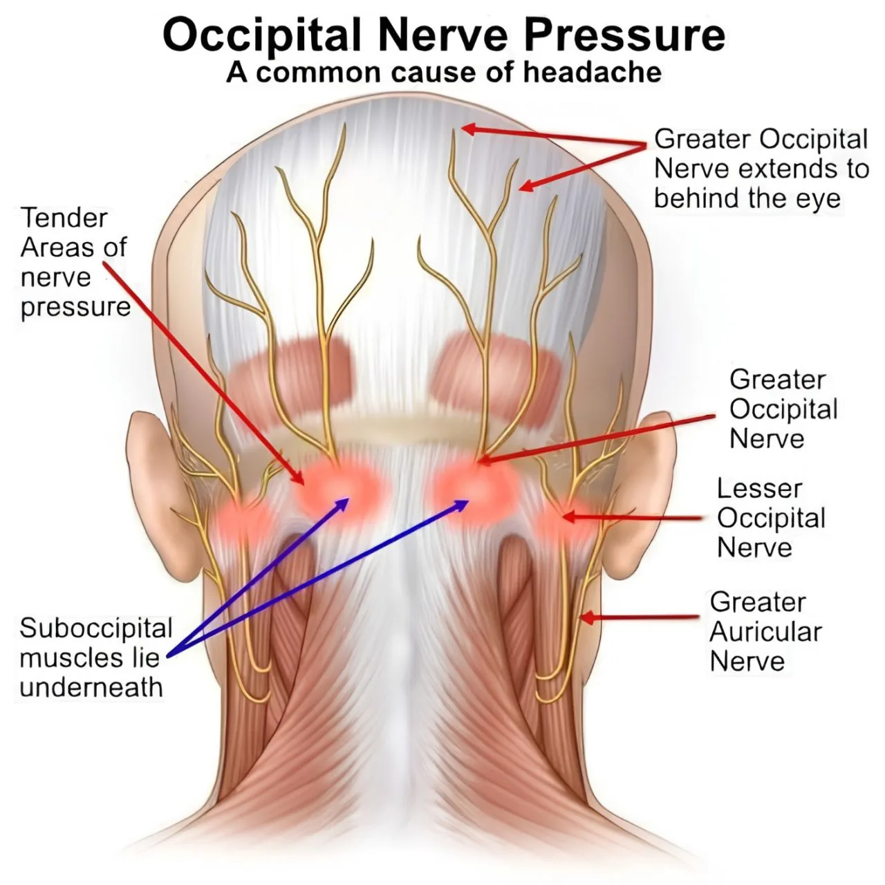
Rectus capitis posterior minor connects through a gap in the bone to the dura mater that wraps your brain and spinal cord. The neck and the head were never two separate problems. Here is how a cervicogenic headache behaves, the one-minute test that identifies it, why it gets felt behind an eye, and the trial evidence that it responds to treatment.
A Muscle at the Base of Your Skull Is Attached to the Covering of Your Brain
There is a small muscle at the base of your skull called rectus capitis posterior minor. It is one of the suboccipital group - the deep layer that sits underneath the big neck muscles you can actually feel.
Anatomists dissecting the back of the neck found something that changed how this region is understood: a band of connective tissue running from that muscle, through a gap in the bone, attaching directly to the dura mater - the membrane that wraps your brain and spinal cord.
It is called the myodural bridge. It is a real, dissectable structure, not a theory.
So the idea that the neck and the head are two separate problems was never really true. There is a mechanical, continuous connection between a muscle you can put your thumb on and the covering of your central nervous system.
Which matters, because a large number of headaches are coming from the neck and being treated as though they are not.
How a Neck-Driven Headache Actually Behaves
Cervicogenic headache has a recognisable pattern. It is not subtle once you know what to look for.
- It starts at the base of the skull and travels forward, often to behind one eye
- It stays on the same side. It tends not to swap sides between attacks the way migraine often does
- There is usually a tender spot back there that, when pressed, reproduces your actual headache rather than just hurting locally - this is the single most useful clue
- It is triggered by sustained head position. Which is why it arrives after driving, a long stretch at a desk, or a night on a bad pillow
That third point deserves emphasis. Local tenderness is common everywhere. Referred reproduction - pressing one spot and feeling your familiar headache switch on somewhere else - is a different finding entirely, and it points at the neck.
If your headache has all four features, the probability that your neck is involved is high enough that it should be assessed properly rather than absorbed for another decade.
There Is a One-Minute Test For It
The cervical flexion-rotation test isolates the joint between your first two vertebrae - the C1-C2 segment, which supplies roughly half of all rotation in your neck.
The neck is bent fully forward, which locks the segments below. Then the head is rotated to each side, and the available range is measured.
The numbers:
- Normal: around 44 degrees to each side
- In one study, people with headache coming from the neck averaged 25 degrees
- People with migraine averaged 42 degrees - essentially normal
- Under 30 degrees is treated as a positive finding
That separation is what makes the test useful. It does not merely show that a neck is stiff; it discriminates between two conditions that present in a very similar way to the person experiencing them.
A physiotherapist can perform it in about a minute. If you have had a one-sided headache from the base of your skull for years and nobody has ever measured this, that is a genuine gap in your assessment - not a reflection on you.
Why the Neck Is Felt Behind the Eye
This is the piece that explains the misdiagnosis, and it is pure neuroanatomy.
Sensory nerves from your upper cervical spine (C1-C3) feed into the same relay nucleus in the brainstem as the trigeminal nerve, which carries sensation from your face and forehead. That shared relay is called the trigeminocervical complex.
Two different sources arrive at the same second-order neurons. Downstream, the brain has no separate label for "this came from the neck" versus "this came from the forehead" - it only receives the output of cells that both inputs converge on.
The consequences are directly observable:
- Pressure at the back of the skull is felt behind an eye
- An anaesthetic injection into the back of the head can settle pain at the front of it
- A neck problem produces a symptom pattern that looks, to the patient, exactly like a primary headache disorder
This is convergence, the same mechanism behind cardiac pain in the left arm. The location you feel is a best guess by a system with limited resolution - not a map reference.
Those Little Muscles Are Sensors, Not Engines
The suboccipital muscles are not there for strength. They are tiny and mechanically weak, and they were never doing the heavy work of holding your head up.
What they are is densely packed with proprioceptors - muscle spindles reporting position and movement - at up to a hundred times the density found in your trapezius.
This region is one of the most information-rich pieces of your musculoskeletal system. It tells your brain where your head is in space, and that signal is integrated with your vestibular system and your eyes to keep the world stable while you move.
Which explains a set of symptoms that otherwise seem unrelated to a neck:
- Dizziness and unsteadiness that is not vertigo and not cardiac
- Visual strain - focusing feels effortful, screens become intolerable
- A vague "not quite here" feeling that is difficult to describe to a clinician
When the sensory report from your upper neck disagrees with your eyes and inner ear, your brain has to reconcile a conflict. Dizziness and visual fatigue are the cost of that reconciliation, not separate problems.
The Useful Part: This Responds to Treatment
This is not a diagnosis of exclusion you get handed and then carry.
In a randomised trial of 200 people with neck-driven headache, both specific neck exercise and manual therapy significantly reduced headache frequency and intensity - and the improvement was still present at twelve months.
Twelve-month durability is unusual and important. It suggests the changes are structural and functional in the tissue and the control system, not a short-lived treatment effect.
What that treatment tends to look like in practice:
- Low-load deep neck flexor training - endurance, not strength; small ranges, many repetitions
- Restoring C1-C2 rotation specifically, rather than generic "neck stretches"
- Manual therapy to the upper cervical segments, used to open a window for the exercise to work in
- Load management for sustained positions - the fix is usually frequency of position change, not the perfect ergonomic chair
- Addressing sleep position and pillow height, because eight hours of sustained end-range is a large daily dose
Not every headache is a neck headache. Migraine is real, common and needs its own management. But if yours starts at the base of your skull, sits on one side, and has a spot you can press that recreates it, that is worth investigating properly.
💜 You have a structure at the base of your skull connected to the covering of your brain, packed with more position sensors than almost anywhere else in your body. It is not a surprise that it can generate headaches. It is a surprise how rarely anyone looks.
Recalibrate is an education and self-tracking tool, not a diagnostic service or a replacement for medical care.
Related articles
- The Ache Two Days After a Hard Workout Is Not Coming From Your Muscles
Sixteen people were injected with the same irritant twice - once into muscle, once into the fascia on top of it. The fascia injection hurt 86% more at peak, lasted up to twice as long and spread across an area 65% larger. No standard scan assesses this tissue. Here is what fascia actually is, why foam rolling does not do what you were told, and what genuinely changes it.
- Your Brain Has a Volume Control for Pain. In Chronic Pain, It Can Get Stuck Turned Up.
Pathways run from your brainstem into your spinal cord and set how much of a signal gets through. They can suppress it - which is why an athlete finishes the match and finds the injury afterwards. The same circuitry can amplify. Here is the neuroscience of descending facilitation, central sensitisation, and why understanding the mechanism is itself one of the best-evidenced treatments we have.
- Leveraging Neuroplasticity in Chronic Pain Recovery
How chronic pain recovery can use neuroplasticity through education, graded exposure, attention training, regulation and repeated safety signals.